


Eye and Retina: cell therapy for retinal damage of traumatic or vascular origin.
The retina is the neurosensory tissue that captures light and converts it into information the brain interprets as vision. When its structure is acutely damaged by trauma or a disruption in blood flow, visual function can be compromised. Even after the initial eye emergency has been resolved, areas of structural damage that do not recover on their own may remain. At LYRACELL we assess the condition of the retina and the affected layers to determine whether cell therapy can be part of the approach to retinal damage of traumatic or vascular origin.
Personalized cell therapyComprehensive medical evaluationIntravenous administrationIn-house laboratory
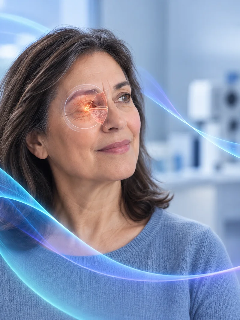
Understanding the retina
The retina is made up of multiple layers of specialized nerve cells, including photoreceptors, bipolar cells and ganglion cells, which work together to capture light and transmit visual information to the brain through the optic nerve.
Because of its characteristics, the retina is considered an extension of the central nervous system. Its function also depends on a precise blood supply and on structures that maintain a protected environment for its cells.
When trauma shifts its position or an interruption in blood flow compromises its blood supply, retinal cells can begin to deteriorate.
Traumatic retinal detachment
A blow or injury to the eye can cause the retina to separate from the structures that provide it with support and nourishment.
When that contact is lost, the supply of oxygen and nutrients to the neural layers can be interrupted. That is why retinal detachment is an eye emergency that requires immediate care and treatment.
Even after surgical repair, structural damage may remain in certain layers of the retina.
Vascular damage to the retina
A blockage of a retinal artery, a retinal vein or one of their branches can disrupt blood flow to a specific region of the retina.
Reduced or interrupted blood supply causes ischemia and can damage the nerve cells in the affected area. Once the acute event is under control, studies make it possible to assess what damage remains and how much viable tissue is preserved.
The remaining damage also matters
Cell therapy does not replace urgent treatment of a retinal detachment or an acute vascular event.
This approach is evaluated afterward, once the initial emergency has been treated and measurable structural damage and impaired visual function persist.
This indication is specifically intended for acute or subacute damage of traumatic or vascular origin in a previously healthy retina. It is not intended for chronic degenerative processes such as maculopathy.

How can it present?
Loss of visual function
Damage to the neural layers can affect vision to varying degrees, depending on its location and extent.
Visual field changes
Certain areas of vision may be affected depending on the region of the retina involved.
After-effects of trauma
Even after the initial eye injury has been resolved, visual impairment related to the remaining retinal damage may persist.
After-effects of a vascular event
An interruption in blood flow can leave areas of neural damage even after the acute episode is under control.
A therapy directed at the retinal tissue that remains damaged after the initial event.
Unlike other tissues, the retina has a very limited capacity for spontaneous regeneration. When trauma or a vascular event causes the loss of retinal cells, the damage may persist even after the initial cause has been resolved.
In these cases, cell therapy aims to support biological processes related to the repair and remodeling of the retinal environment and to support the recovery of preserved visual function.
The treatment used at LYRACELL combines two components:
- Stem cells obtained from adipose tissue: processed and prepared in the laboratory and directed toward cell types linked to the retina and to nerve and vascular tissue.
- Immune system cells obtained from the blood: take part in preparing the cell therapy and directing it toward the structures being treated.
Under the protocols of the Moviglia Method, the LYRACELL team prepares a specific cell combination according to the origin, location and extent of the damage identified.
The combination of both cell types aims to support the body’s natural repair processes through a personalized approach and under medical and safety criteria.
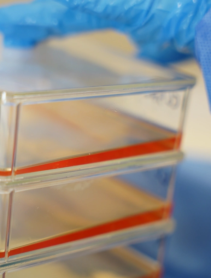
From the laboratory to the retina
The stem cells are obtained from the patient and, together with the immune system cells, are processed in the laboratory following the protocols of the Moviglia Method. Because of the particular characteristics of the retina and the barriers that protect the nervous system, the protocol includes a sequential intravenous administration in two stages: first, the specific immune system cells are administered and, forty-eight hours later, the cells previously prepared and directed toward cell types linked to the retina and nerve tissue are infused. This strategy aims to support the interaction of the cell preparation with the affected regions without requiring an invasive application directly into the eye.


Four stages to address the retinal damage that remains after the initial injury.
The treatment is defined according to the origin of the damage, the layers involved, its extent and the presence of viable neural tissue.
1
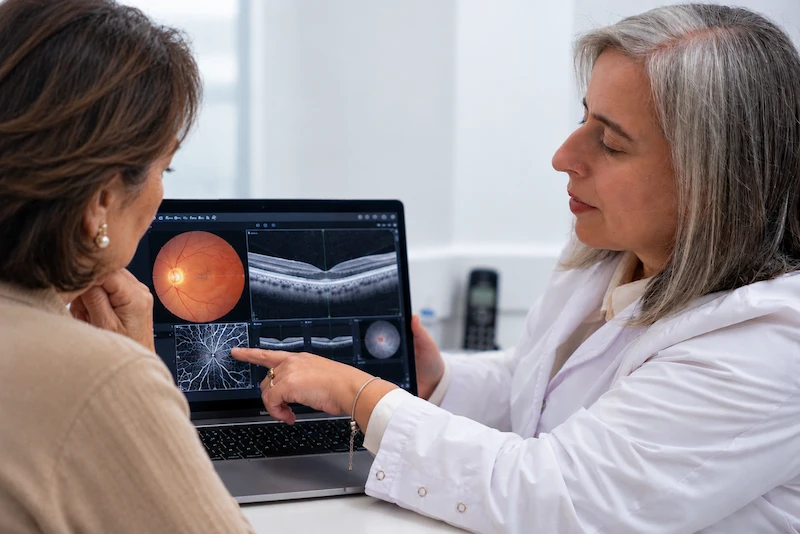
Evaluation
The medical history, the mechanism of injury —traumatic or vascular— and the medical evaluation are reviewed, in coordination with your treating specialist. This is complemented with optical coherence tomography —OCT—, angiography and fundus examination to define the location and extent of the damage.
2
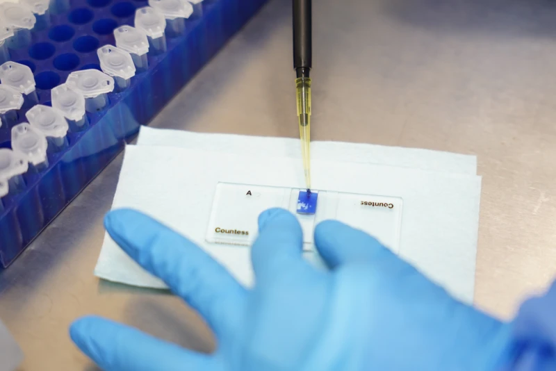
Treatment proposal
The medical team assesses how much viable retinal tissue remains and whether the patient is eligible, and defines the cell volume and the direction of the preparation. Adipose tissue and peripheral blood are collected, and a formula adapted to the tissue to be treated is prepared in the laboratory.
3
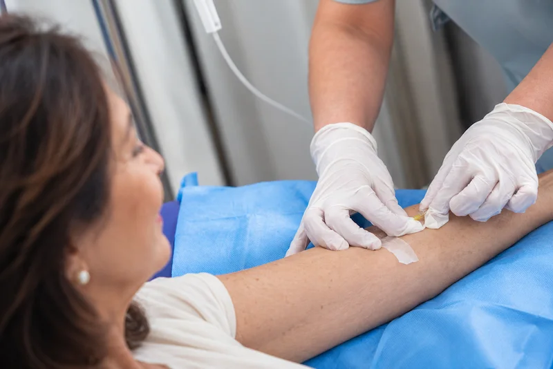
Application
Administration is intravenous and in two stages: first the specific immune system cells are infused, which direct the therapy toward the affected regions, and 48 hours later the stem cells prepared for the retina and nerve tissue. The frequency depends on the severity and progress.
4
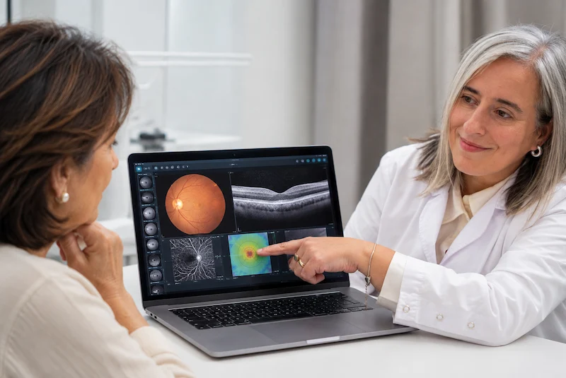
Follow-up
Progress is monitored with serial retinal imaging studies and vision assessments, and eye movement and visual field rehabilitation may be added. Two or three applications over 6 to 12 months may be planned, with final studies to compare with the starting point.
An accurate evaluation makes it possible to treat the specific problem, not just the symptom.
Could this treatment be an option for you?
This indication is specifically intended for patients with loss of visual function secondary to acute or subacute retinal damage of traumatic or vascular origin, once the initial eye emergency has been resolved.
It may be an option if…
You have had a traumatic retinal detachment and structural damage persists after the initial treatment.
You have had an acute retinal vascular event with visual after-effects.
There is loss of visual function related to the damage identified.
Studies show objective structural changes in the layers of the retina.
The initial eye emergency has already been resolved with the corresponding medical or surgical treatment.
Studies show remaining viable neural tissue suitable for this approach.
Another approach may be needed if…
There is a retinal detachment that still requires urgent eye care.
The condition is a maculopathy or another chronic degenerative retinal process.
There is a progressive retinal degeneration of genetic origin.
There is complete, long-standing retinal atrophy, with no signs of remaining viable neural tissue.
The vision loss is due to a refractive error.
The origin lies in a proximal lesion of the optic nerve or in the visual pathway in the brain.
Retinal studies do not explain the vision loss and other possible causes need to be investigated.
The indication is defined individually based on the origin of the injury, the studies and the presence of viable retinal tissue.
Images that make it possible to compare progress.
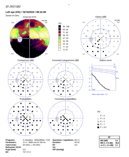
Follow-up images make it possible to compare the visual field before treatment and at later check-ups. They correspond to specific cases: the response, timing and degree of progress vary from patient to patient.
Before
After
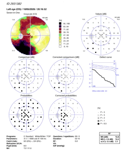
A 72-year-old patient with a history of herpetic neuritis that, in this case, damaged the optic nerve. The patient received 2 regenerative treatments directed at the retina and the optic nerve. Comparing their visual field before (October 2024) and after (May 2026), we found:
- 68% more visual sensitivity: the eye’s ability to perceive light went from 7.6 to 12.8 dB.
- 28% less vision loss: the mean defect dropped from 18.8 to 13.5 dB.
- 40% less generalized damage: the diffuse defect decreased from 13.3 to 8.0 dB.
- Smaller areas without vision: the study map shows fewer black areas, especially in the upper and central parts.
These results show a measurable improvement in this patient’s vision.
Results may vary from person to person. Each case is evaluated individually.
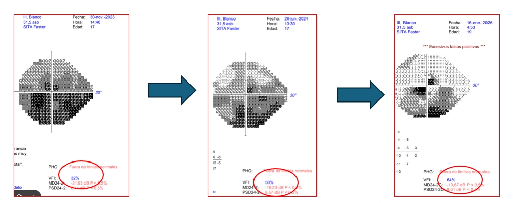
A 19-year-old patient diagnosed with a retinal detachment that occurred while playing sports.
The patient received 3 applications of regenerative treatment between the first and second study, and 6 more treatments between the second and third. Comparing their visual fields over two years, we found:
- Their visual function doubled: the visual function index went from 32% to 64%.
- 38% less vision loss: the mean defect dropped from -21.93 to -13.67 dB.
- Improvement at every stage: after the first 3 applications, vision loss decreased by 17%. With the following 6 treatments, it dropped a further 25%.
- Smaller areas without vision: in the study maps, the dark areas (where the patient could not see) gradually shrink, especially in the lower half of the visual field.
These results show a progressive, measurable recovery of vision that continues to improve as treatment advances.
Results may vary from person to person. Each case is evaluated individually.
Treating the tissue is part of the process. Assessing how vision responds is too.
When treating the retina, progress needs to be observed from both a structural and a functional point of view. That is why follow-up combines imaging studies, vision assessments and a rehabilitation plan adapted to each patient’s needs.
Retinal follow-up
Serial studies make it possible to observe how the retinal layers evolve and to compare the findings with the images obtained before treatment.
Visual rehabilitation
Exercises aimed at retraining eye movement and training the visual field can be part of the rehabilitation process when indicated.
The program is adapted to the type and extent of each patient’s visual impairment.
Measurable results
Progress can be assessed through optical coherence tomography and other retinal imaging studies, together with functional vision tests.
This makes it possible to compare the changes observed during treatment with the starting point.
Frequently asked questions
The retina is a neurosensory tissue made up of different layers of specialized cells that capture light and transmit visual information to the brain through the optic nerve.
No. A retinal detachment is an eye emergency and must first receive the corresponding treatment. Cell therapy is evaluated afterward, when structural damage and impaired visual function persist.
This indication is specifically intended for acute or subacute damage of traumatic or vascular origin in a previously healthy retina.
No. Maculopathy and other chronic or progressive retinal degenerations are not part of this indication, since they have different mechanisms of damage and tissue characteristics.
The evaluation may include optical coherence tomography —OCT—, angiography and fundus examination, in addition to the corresponding medical evaluation.
The base treatment is autologous: the stem cells are obtained from the patient’s own adipose tissue and the immune system cells from their blood. In certain cases, depending on the medical indication, immune system cells from a selected donor may be used.
Under this protocol, no. Administration is intravenous and in two stages, without applying the preparation directly into the eye.
The protocol first administers specific immune system cells and, 48 hours later, infuses the previously prepared cells. This sequence aims to help direct the cell therapy toward the affected regions.
Usually, two to three applications over a period of 6 to 12 months may be planned. The number is defined at the start of treatment, according to the extent of the damage, as part of a plan with specific goals. At the end of the plan, progress is assessed and, if needed, further interventions are defined.
Serial retinal imaging studies are combined with functional vision assessments to compare progress with the initial state.
The first step is understanding what damage remains in the retina.
Every treatment begins with a medical evaluation, in coordination with your treating specialist, to determine the extent of the injury, the layers involved and the presence of viable neural tissue, and to establish whether this approach may be right for your case.