


Ankle: cell therapy applied to osteoarthritis and tendon injuries.
Ankle pain can affect everyday activities such as standing, walking, running or jumping. It may appear suddenly after a twisting sprain, a fracture or another traumatic injury, or develop gradually. At LYRACELL we evaluate the affected structures to identify the source of the pain and define a personalized treatment.
Personalized cell therapyUltrasound-guidedMinimally invasiveIn-house laboratory

Understanding the ankle
The ankle is formed by the junction of three bones —tibia, fibula and talus—, protected by cartilage and stabilized by a complex system of lateral and medial ligaments. Tendons such as the Achilles tendon, the peroneal tendons and the posterior tibial tendon take part in movement and load distribution.
Pain may be mainly associated with:
Joint wear and damage
Progressive deterioration of the cartilage can lead to ankle osteoarthritis. There may also be bone edema, osteochondral lesions of the talus and irregularities such as osteophytes or impingement, which cause friction, pain and limited movement.
Ligament instability
Sprains, micro-tears or complete tears of the ligaments —common after poorly healed sprains— can compromise ankle stability, make new twists more likely and speed up joint wear.
Tendon injuries
Tendinopathies, wear or tears of the Achilles tendon or the peroneal tendons can alter how force is distributed when walking or jumping and contribute to chronic inflammation, atrophy and loss of muscle strength.
In many cases, several structures are affected at the same time.
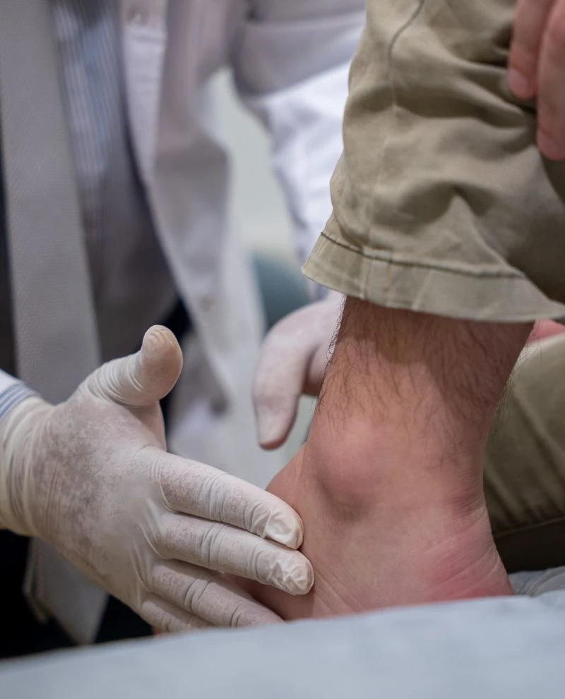
How can it present?
Pain when standing or walking
Discomfort may appear when putting weight on the foot, walking, running or jumping, or after standing for a long time.
Instability or frequent sprains
Ligament involvement can make you feel unsteady when putting weight on the foot and make new sprains more likely.
Inflammation or swelling
Edema may appear around the ankle, especially after activity.
Stiffness or loss of mobility
Bending or straightening the ankle may become difficult or painful, for example when going up or down stairs.
Loss of strength or limits in sports
Tendon injuries can reduce push-off when walking, running or jumping, and limit daily or sports activities.
Your own cells, directed toward the tissue that needs treatment.
The body has natural self-repair mechanisms. However, in the case of advanced structural injuries, tendon tears, severe sprains with chronic instability or continuous micro-injuries caused by improper weight-bearing, that capacity may not be enough.
In these cases, the contribution of cells seeks to support biological processes related to tissue repair and remodeling, modulate inflammation and support the recovery of ankle function.
The personalized treatment combines two key components: stem cells, which are part of a preparation intended to support biological processes related to tissue repair and remodeling, and immune system cells, which help direct the stem cells toward the tissue that needs treatment —cartilage, tendon, ligament or bone—.
Under the protocols of the Moviglia Method, the LYRACELL team obtains stem cells from the patient’s own fat. These are processed and expanded in the laboratory and then combined with tissue-specific immune system cells. The preparation includes pre-differentiated stem cells and immune system cells directed toward the structures identified in the diagnosis.
The combination of both cell types seeks to support the body’s natural repair process, through a personalized approach and under strict safety protocols.
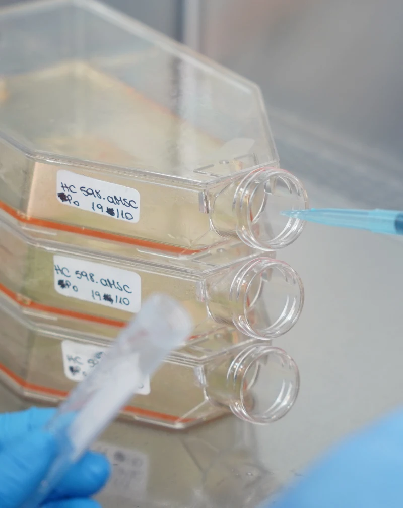
From the laboratory to your ankle
The cells are processed in the laboratory and prepared according to the tissue identified in the evaluation before the targeted application.
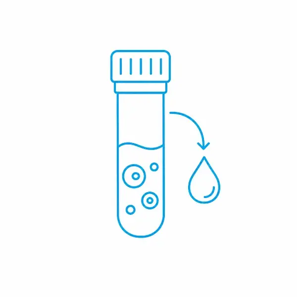


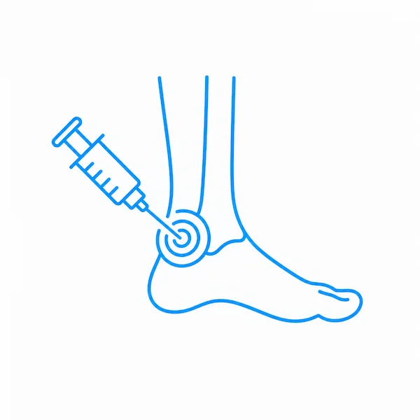
Four stages, one process defined for each case.
From the initial evaluation to the follow-up of your progress, each stage of the treatment is defined by the medical team according to your case.
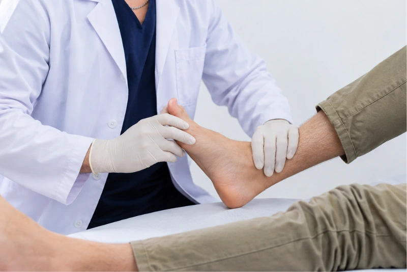
1
Evaluation
We analyze the medical history, any history of sprains, trauma or sports practice, and imaging studies —X-rays, ultrasound, CT or MRI— to identify the affected structures and determine the degree of the injury.
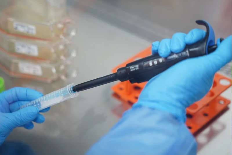
2
Treatment proposal
If the patient is eligible, a plan is designed according to the extent of the injury and the tissues involved —cartilage, ligaments, tendons or bone—. Adipose tissue and peripheral blood are collected, and the cell combination adapted to the affected tissue is prepared in the laboratory.
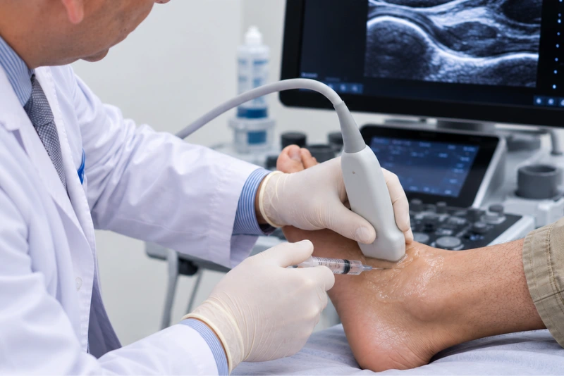
3
Application
In a minimally invasive way and under ultrasound guidance, the cells are applied into the joint, the bone or the affected ligaments and tendons. If arthroscopy, ligament repair or other surgery is required, they can be applied during or after it. The number of applications depends on the case.

4
Follow-up
At first there may be local discomfort or inflammation. Progress is evaluated at later medical check-ups and can be supported with rehabilitation to strengthen the peroneal and tibial muscles, work on proprioception and balance, and restore mobility. Clinical, functional and imaging check-ups follow.
An accurate evaluation makes it possible to treat the specific problem, not just the pain.
Could this treatment be an option for you?
The indication is defined individually based on the medical evaluation, symptoms and imaging studies.
It may be an option if…
You have persistent pain, instability or functional limitation in the ankle.
You have had recurrent sprains or feel unsteady when putting weight on the foot or moving.
Your medical studies show a structural change.
The loss of stability affects your daily or sports activities.
You want to preserve ankle function and act on signs of deterioration.
Another approach may be needed if…
There is very advanced osteoarthritis, severe or total loss of joint space, rigid bone deformity or continuous friction. These conditions may require joint fusion or a joint replacement.
The pain or numbness has a peripheral neurological origin, such as tarsal tunnel syndrome.
The symptoms come from compression of the nerve roots in the lumbar spine.
The evaluation makes it possible to distinguish a structural problem in the ankle from other causes that can produce similar symptoms.
Regaining stability also means trusting movement again.
The recovery plan may include rehabilitation to strengthen the stabilizing muscles, work on proprioception and balance, and gradually restore range of motion. Follow-up makes it possible to assess how the ankle responds to weight-bearing, loads and physical activity.
First days
There may be transient local discomfort or inflammation. The team indicates the necessary care and any temporary adjustments to activity and weight-bearing.
Rehabilitation and progress
Progress is evaluated at later medical check-ups. Rehabilitation supports this process by strengthening the peroneal and tibial muscles, working on proprioception and balance, and restoring range of motion.
Measurable results
Clinical and functional check-ups make it possible to assess pain, stability, strength and function. When indicated, new imaging studies are added to compare progress with the starting point.
Frequently asked questions
It depends on the diagnosis. Osteoarthritis, ligament instability, osteochondral lesions of the talus and tendon injuries are evaluated to determine whether cell therapy can be part of the approach.
It is one of the most common reasons for consultation. The indication depends on the degree of ligament instability and its impact on function, and is defined after a medical evaluation.
The base treatment is autologous: the stem cells are obtained from the patient’s own adipose tissue and the immune system cells from their blood. In certain cases, depending on the medical indication, immune system cells from a selected donor may be used.
The medical team analyzes your medical history, any history of sprains or trauma, and imaging studies such as ultrasound, X-rays or MRI.
Yes. The cells are applied under ultrasound guidance into the joint space, the bone, or the affected ligament or tendon, depending on where the damage is located.
In some cases it can be an alternative to surgery. If the injury requires arthroscopy or ligament repair, the cells can be applied during or after the procedure as a complement, not a replacement.
It depends on the type of injury and each patient’s progress. The team gives specific care instructions for the first days and then defines a gradual recovery plan.
The return is gradual. It depends on clinical progress, proprioception and balance work, and the medical team’s instructions. There is no single timeline.
Through clinical check-ups and a functional assessment of pain, stability and strength. When appropriate, imaging studies are also used. The results are compared with the starting point.
If the diagnosis points to another approach, such as surgery or a neurological evaluation, the team will tell you and guide you on the next steps. This can happen, for example, if the symptoms are due to tarsal tunnel syndrome or compression in the lumbar spine.
It is a developing field of research. Results vary depending on the type of injury and the protocol used. That is why each indication starts from an individual diagnosis and is accompanied by follow-up with measurable parameters.
The first step is understanding your case.
Every treatment begins with a medical evaluation to understand your diagnosis, identify the affected structures and determine which approach may be right for you.