


Jawbone: cell therapy applied to loss of bone density and volume.
The maxillary bone supports the teeth and implants and, together with the zygomatic (cheek) bone, helps support the tissues of the face. When it loses density, height or volume, it may become difficult to hold implants or prostheses, chewing may become uncomfortable and the facial structure may gradually change. At LYRACELL we evaluate the condition of the maxillary and zygomatic bone to determine the degree of bone loss and establish whether cell therapy can be part of a personalized treatment.
Personalized cell therapyLocalized applicationCT follow-upIn-house laboratory

Understanding the jawbone
The maxillary bone and the malar or zygomatic bone are part of the central structure of the face. They support the teeth and implants, contribute to the projection of the cheekbones and support the soft tissues of this region.
After tooth loss, an infection, trauma or surgery, or as part of aging, the bone tissue can begin to lose density, height and volume.
This deterioration can occur at different levels and, often, several of them appear at the same time.
Bone resorption and ridge atrophy
After a tooth is lost, the bone stops receiving part of the mechanical stimulus that helped maintain its structure.
Over time, this can lead to a progressive reduction in the width and height of the bone ridge. A history of infections, trauma and other processes that can affect the tissue may also play a role.
Loss of density and structural quality
When the natural balance of bone remodeling is disrupted, its internal trabecular structure can decrease.
The tissue becomes less dense and may lose its ability to provide adequate support for teeth, implants and soft tissues.
Loss of midface support
Deterioration of the zygomaticomaxillary structure can also change the support of the soft tissues.
As bone volume decreases, changes in the facial contour may occur, such as less projection of the cheeks or deeper folds in certain areas.

How can it present?
Unstable implants or prostheses
The quantity and quality of the available bone are key factors in providing adequate support.
Difficulty or discomfort when chewing
Structural loss can alter the function of the area and affect everyday activities.
Progressive loss of bone volume
Imaging studies may show a decrease in the height, width or density of the bone.
Changes in facial support
Maxillary and zygomatic bone loss can affect the soft tissues and gradually change the contour of the face.
A therapy aimed at strengthening the remaining bone and supporting its remodeling.
Bone tissue has natural remodeling and repair mechanisms. However, in the case of significant atrophy, long-standing tooth loss, chronic infections or certain processes associated with aging, those mechanisms may not be enough to address the existing deficit.
In these cases, cell therapy seeks to support biological processes associated with bone matrix remodeling, local vascularization and the strengthening of the remaining tissue.
The treatment used at LYRACELL combines two components:
- Stem cells obtained from adipose tissue: they are processed and pre-differentiated in the laboratory toward progenitors linked to bone tissue and the vascular component.
- Immune system cells obtained from the blood: they take part in preparing and directing the therapy toward the areas to be treated.
Under the protocols of the Moviglia Method, the LYRACELL team prepares a specific cell combination according to the location and extent of the identified deficit.
The combination of these components seeks to support the body’s natural repair processes through a personalized approach and under medical and safety criteria.
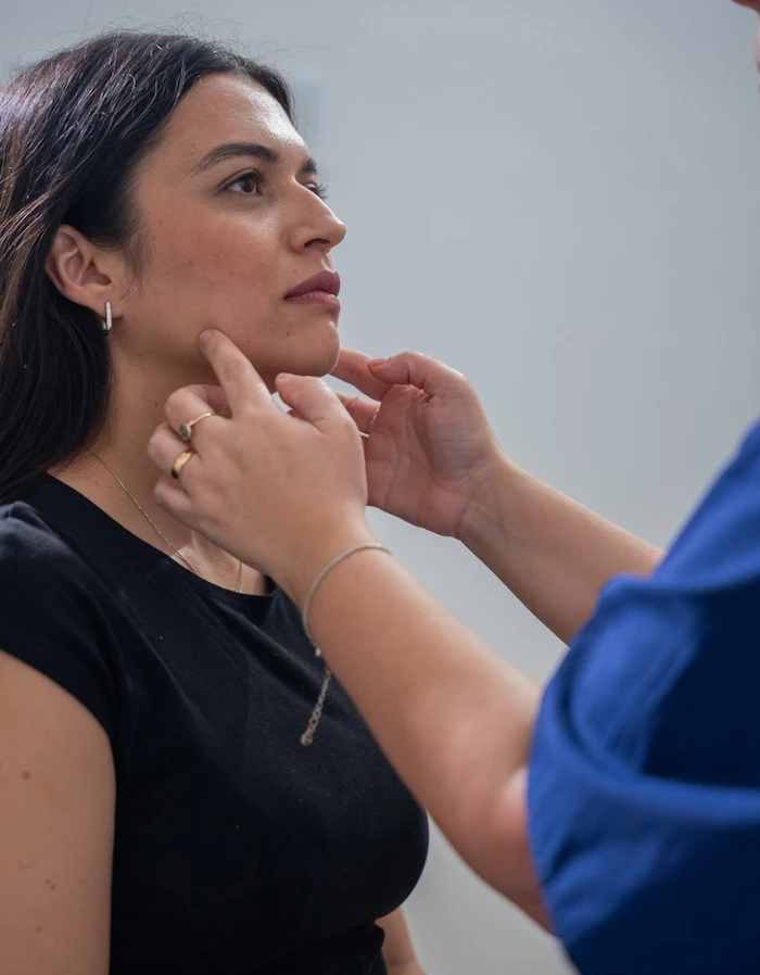
From the laboratory to the jawbone
The stem cells are obtained from the patient and, together with immune system cells, are processed in the laboratory following the protocols of the Moviglia Method. Based on the diagnosis, a cell formula adapted to each patient’s bone deficit is prepared. The application is mainly local and is performed through micro-injections into the remaining bone and the tissues of the affected maxillary or zygomatic regions. When the goal is not only to improve the quality of the existing bone but also to generate more volume, the cell treatment can be combined with a bone matrix that provides a scaffold in the area with the greatest deficit.



Four stages to address bone deficit according to its extent and location.
The treatment is planned based on the quantity and quality of the available bone and, when appropriate, in coordination with the treating dentist.
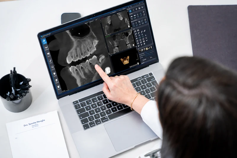
1
Evaluation
We review the history of tooth loss, infections, surgeries and trauma, and how the atrophy has progressed. This is complemented with cone beam computed tomography —CBCT— and bone density analysis to determine the location and extent of the deficit.
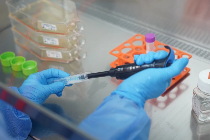
2
Treatment proposal
We determine how much bone remains, its quality and which areas to treat, together with the dentist if implant planning is involved. A local cell application may be indicated or, if volume is needed, combined with a bone matrix. If the patient is eligible, adipose tissue and blood are collected.
3
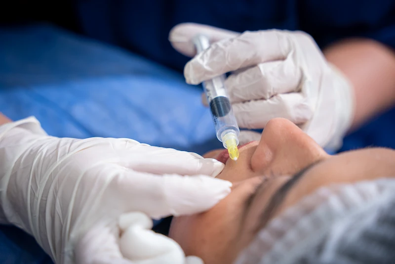
Application
The cells are applied through local micro-injections into the remaining bone and the affected maxillary or zygomatic regions and, if volume is needed, together with the implanted matrix. The number and frequency depend on the deficit, the dental strategy and progress.

4
Follow-up
At first there may be local discomfort or inflammation. Progress is evaluated over weeks and months with clinical and CT follow-up of bone density, quality and, if a matrix was used, volume. If there is dental rehabilitation, the biomechanical load is monitored with the treating professional.
An accurate evaluation makes it possible to treat the specific problem, not just the symptom.
Could this treatment be an option for you?
This approach can be evaluated in people with resorption, atrophy or loss of density of the maxillary or zygomatic bone, as long as there is a structural deficit that can be confirmed by imaging studies.
It may be an option if…
You have bone loss after a tooth extraction.
There is atrophy or resorption of the maxillary ridge.
Studies show loss of bone density, height or volume.
There are bone sequelae after certain trauma or surgeries.
The bone deficit compromises the planning of implant or prosthetic treatment.
The quality of the remaining bone needs to be improved.
There is a volume deficit for which a strategy combined with a bone matrix has been indicated.
The loss of maxillary or zygomatic bone support is affecting the facial structure.
Another approach may be needed if…
There is severe osteonecrosis of the jaw that requires prior surgical treatment.
There is extensive bone destruction associated with a severe active infection.
Extensive surgical debridement is needed before considering a regenerative strategy.
There is a neoplasm in the maxillofacial region that has not yet received the appropriate treatment.
A destructive bone lesion is identified that first requires specific oncological or surgical treatment.
The indication is determined based on the clinical evaluation, imaging studies and the characteristics of each patient’s bone deficit.
Treating the bone is part of the process. So is monitoring how it responds to loads.
The jawbone is subject to constant forces during chewing and, in many patients, is part of a broader dental treatment plan. That is why follow-up is not limited to the moment of the cell application.
First days
There may be transient local discomfort or inflammation in the treated areas. The medical team provides the corresponding follow-up and indicates the necessary care for each procedure.
Joint follow-up
When there is implant or prosthetic planning, LYRACELL works in coordination with the treating dentist to follow the progress of the bone and gradually monitor the biomechanical load.
Measurable results
CT follow-up makes it possible to compare the progress of the tissue with its initial condition and assess parameters such as density, structural quality and, when appropriate, bone volume.
These results can also help define the next steps of the dental or prosthetic treatment.
Frequently asked questions
After a tooth is lost, the bone stops receiving part of the mechanical stimulus associated with that tooth. Over time, the width and height of the bone ridge can progressively decrease.
No. It can be evaluated for resorption, atrophy or loss of density of the maxillary or zygomatic bone. Implant or prosthetic planning is one of the situations in which it can be part of a joint strategy.
The evaluation may include cone beam computed tomography —CBCT—, X-rays and bone density analysis, in addition to the corresponding clinical examination.
The base treatment is autologous: the stem cells are obtained from the patient’s own adipose tissue and the immune system cells from their blood. In certain cases, depending on the medical indication, immune system cells from a selected donor may be used.
The application is mainly local and is performed through micro-injections into the remaining bone and the affected areas.
When the goal requires generating more volume, the protocol includes combining cell therapy with a bone matrix that provides the necessary scaffold. The strategy depends on the extent and characteristics of the deficit.
Yes. When there is implant or prosthetic planning, the treatment is defined together with the treating dentist and is accompanied by follow-up of the bone’s progress and the biomechanical load.
Mainly through clinical evaluation and CT follow-up, comparing the density, structural quality and, when appropriate, the volume of the bone with the initial studies.
The first step is knowing how much bone remains and what it needs to recover.
The evaluation makes it possible to determine the density, volume and structural quality of the maxillary or zygomatic bone, and to define whether cell therapy, alone or combined with a bone matrix, can be part of the right strategy for your case.