


Trigeminal nerve: cell therapy applied to trigeminal neuralgia.
Intense facial pain, a burning sensation or changes in sensation may be related to involvement of the trigeminal nerve. At LYRACELL we evaluate the condition of the nerve branches and the tissues involved to identify the source of the pain and determine whether cell therapy can be part of a personalized treatment.
Personalized cell therapyLocalized applicationMinimally invasiveIn-house laboratory

Understanding the trigeminal nerve
The trigeminal nerve carries sensation from the face, the oral cavity and the cornea to the brain. It also helps control the chewing muscles.
When there is trigeminal neuropathy, the structure and function of its nerve fibers may be altered. The involvement can affect the transmission of sensory signals and cause pain even in response to stimuli that normally should not trigger it.
Demyelination and axonal damage
Loss or damage of the myelin —the layer that protects the nerve fibers— can change the transmission of electrical impulses and generate abnormal pain signals in response to normally painless stimuli, a phenomenon known as allodynia.
Microvascular or structural compression
Repeated contact or pressure from nearby structures or blood vessels can cause mechanical irritation and inflammation of the nerve root.
Central sensitization and micro-inflammation
Persistent nerve damage can maintain an inflammatory environment both peripherally and centrally and help keep the neuropathic pain circuit active.
These mechanisms can occur at the same time and contribute to the persistence of symptoms.
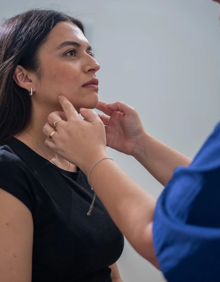
How can it present?
Intense facial pain
It may appear as stabbing episodes or electric shock-like pain in different areas of the face.
Burning or persistent pain
In some cases, the discomfort may feel like burning or continuous pain.
Pain from everyday stimuli
Washing your face, brushing your teeth, talking, chewing or even feeling the wind on your face can trigger pain.
Changes in sensation
Changes in how certain areas of the face or oral cavity feel may appear.
Functional limitation
When symptoms persist, everyday activities such as eating, talking or personal hygiene may be affected.
Your own cells, directed toward the affected nerve tissue.
The body has natural mechanisms to repair its tissues. However, in the case of severe trauma, prolonged mechanical compression or chronic neurological damage, this capacity may not be enough.
In these cases, the contribution of cells seeks to support biological processes related to the support and remodeling of the nerve fiber and its environment, and to support functional progress and the course of neuropathic pain.
The personalized treatment combines two key components:
- Mesenchymal stem cells: they can release neurotrophic factors related to the protection of the axonal structure, remyelination and the remodeling of peripheral nerve tissue.
- Immune system cells: they help direct the preparation toward the cell types linked to the affected tissue and shape its interaction with the inflammatory environment.
Under the protocols of the Moviglia Method, the LYRACELL team obtains stem cells from the patient’s own adipose tissue and immune system cells from the blood. The cells are processed in the laboratory and combined as part of a preparation directed toward the structures of the peripheral nervous system.
The preparation is administered through a localized application around the affected nerve, with the aim of supporting biological processes associated with nerve tissue repair and supporting its clinical and functional progress.
The combination of both cell types seeks to support the body’s natural repair process, through a personalized approach and under strict safety protocols.
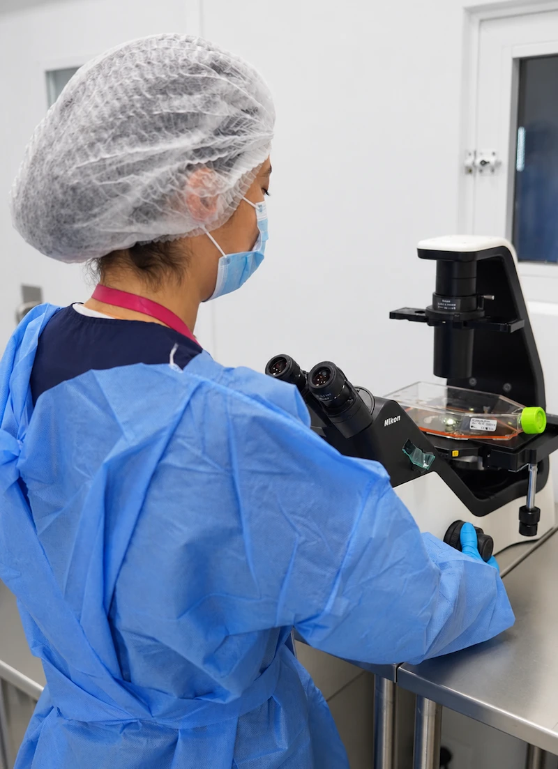
From the laboratory to the trigeminal nerve
The stem cells are obtained from the patient and, together with immune system cells, are processed in the laboratory following the protocols of the Moviglia Method. Based on the diagnosis and the type of involvement identified, the LYRACELL team prepares a specific cell combination before its application around the affected nerve.

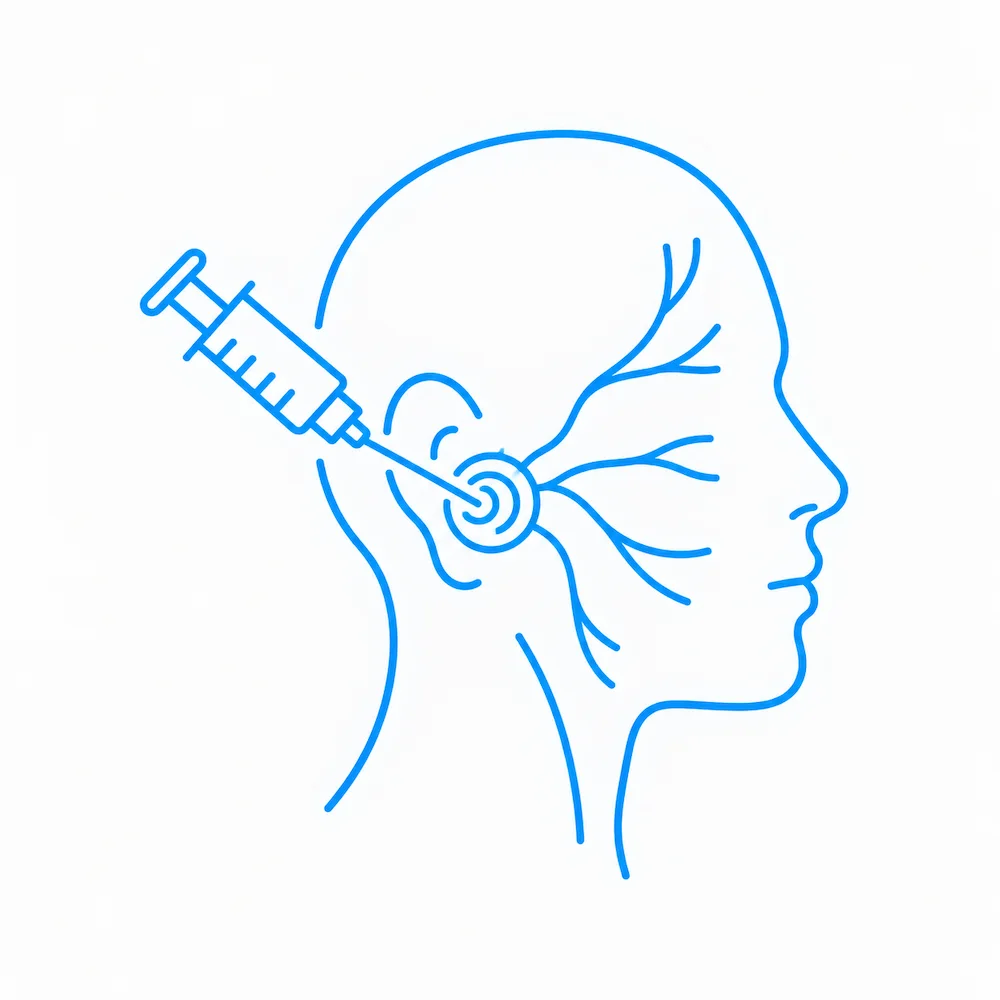
Four stages, one process defined by the source of the pain.
From identifying what is affecting the nerve to following up on its progress, each stage is defined according to the characteristics of each case.
1
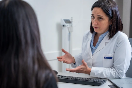
Evaluation
We review the medical history, any history of trauma or previous surgery, and how long the pain has been present. This can be complemented with brain MRI using a cranial nerve protocol, CT and neurophysiological studies to analyze the degree of involvement, identify possible compression and evaluate the surrounding tissues.
2
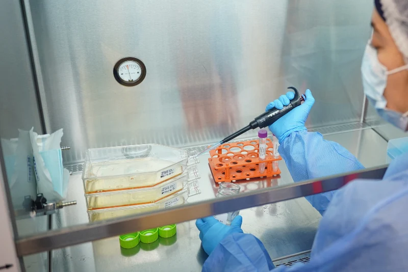
Treatment proposal
The medical team determines whether cell therapy may be appropriate. If the patient is eligible, adipose tissue and peripheral blood are collected, and a cell combination adapted to the needs of the nerve tissue is prepared in the laboratory.
3
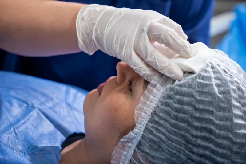
Application
The cell preparation is administered through a localized injection around the affected nerve. The number and frequency of applications are determined individually according to the degree of involvement and the clinical progress observed.

4
Follow-up
Nerve tissue recovery takes time and is evaluated gradually through clinical and functional follow-up. Physical and neurological therapy can complement the treatment to work on desensitizing the area and support the recovery of facial function.
An accurate evaluation makes it possible to treat the specific problem, not just the symptom.
Could this treatment be an option for you?
The indication requires identifying structural or functional involvement of the trigeminal nerve through the medical evaluation and the corresponding diagnostic studies.
It may be an option if…
You have pain associated with trigeminal neuropathy or neuralgia.
You experience burning, sensory changes or pain in response to normally painless stimuli.
The symptoms affect everyday activities such as talking, chewing, washing your face or brushing your teeth.
Studies show measurable structural or functional involvement of the nerve.
There is a history of trauma or surgery related to the nerve involvement.
You want to address the nerve tissue involvement and support the recovery of its function.
You want to act on identified nerve damage before the functional limitation progresses.
Another approach may be needed if…
There is severe tumor compression or a complex vascular malformation that requires neurosurgical intervention or immediate decompression.
The pain has a non-neuropathic origin, such as an acute odontogenic infection or acute sinusitis.
The evaluation identifies another condition that is not linked to the structure or function of the trigeminal nerve.
The indication is defined individually based on the neurological evaluation and the corresponding studies.
Images that make it possible to compare progress.
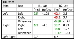
Follow-up studies make it possible to compare the function of the trigeminal nerve before treatment and at later check-ups. They correspond to a specific case: the response, timing and degree of improvement vary from patient to patient.
Before
After
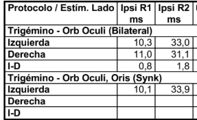
45-year-old patient with left-sided trigeminal neuralgia of 10 years’ duration. In her case, the disease had not responded to any previous treatment: not to medication, radioablation or surgery.
The patient received 3 cycles of regenerative treatment over 12 months. To measure the results, the blink reflex was studied: the trigeminal nerve is gently stimulated and the time it takes the eye to close is measured. When the nerve is damaged, the blink comes late or does not appear.
- Normal blinking restored: before, when the left side of the face was stimulated, the eye-closing reflex was delayed, outside normal values. After treatment, the response returned to normal values and is 22% faster.
- Both sides of the face work the same again: the difference between the affected side and the healthy side was reduced by 79%. Today the left side responds practically the same as the right.
- A nerve that transmits again: these results show that the trigeminal nerve recovered its ability to conduct the signal correctly.
These results are especially significant in a patient who, after 10 years of disease, had not improved with any treatment.
Results may vary from person to person. Each case is evaluated individually.
Nerve tissue has its own timing. Supporting its function is also part of the process.
After the application, the medical team defines the follow-up according to the origin of the involvement, how long it has been present and each patient’s response.
First days
The medical team indicates the post-application care and any temporary changes to activity that apply in each case.
Physical and neurological therapy
Rehabilitation can complement cell treatment with strategies aimed at gradually desensitizing the area and supporting facial function. The plan is adapted to each patient’s characteristics and symptoms.
Measurable results
Follow-up makes it possible to assess changes in the intensity and frequency of pain, sensation and the activities that were limited before treatment. When indicated, new neurological evaluations or complementary studies can be added to compare progress with the starting point.
Frequently asked questions
It is a nerve that carries much of the sensation from the face, the oral cavity and the cornea to the brain. It also helps control the muscles used for chewing.
Damage to the nerve fibers or their myelin layer can alter the transmission of sensory signals. As a result, normally painless stimuli may be interpreted as pain.
The evaluation starts with the medical history and examination and can be complemented with brain MRI using a cranial nerve protocol, CT and neurophysiological or nerve conduction studies.
The base treatment is autologous: the stem cells are obtained from the patient’s own adipose tissue and the immune system cells from their blood. In certain cases, depending on the medical indication, immune system cells from a selected donor may be used.
The cell preparation is administered through a localized injection around the affected nerve, according to the strategy defined for each case.
The number of applications is defined at the start of treatment, according to the degree of involvement, as part of a plan with specific goals. At the end of the plan, progress is evaluated and, if needed, new interventions are defined.
Nerve tissue recovery is gradual and takes time. That is why there is no single timeline: follow-up makes it possible to assess each patient’s clinical and functional changes.
It can be part of the process. Physical and neurological therapy can be used to work on desensitizing the area and to support facial function.
The first step is understanding where the pain comes from.
Every treatment begins with a medical evaluation to identify the origin of the symptoms, understand the condition of the nerve and determine which approach may be right for you.