


Hip: cell therapy for osteoarthritis and trochanteric bursitis.
Hip pain is very common and significantly affects quality of life, walking and the ability to move and play sports freely. It can appear suddenly after a blow or a fall, or develop gradually as a result of wear and certain movement patterns. At LYRACELL we assess the affected structures to identify the origin of the pain and define a personalized treatment.
Personalized cell therapyUltrasound-guidedMinimally invasiveIn-house laboratory

Understanding the hip
The hip is a ball-and-socket joint formed by the head of the femur and the acetabulum of the pelvis. Cartilage protects its surfaces, while the labrum and capsular ligaments provide stability. Muscle groups such as the glutes, iliopsoas and adductors are involved in movement and in distributing loads.
The pain may be mainly associated with:
Joint wear and damage
Progressive deterioration of the cartilage can lead to hip osteoarthritis, or coxarthrosis. Bone edema and irregularities such as osteophytes or femoroacetabular impingement may also appear, causing friction, pain and limited range of motion.
Labral tears
The labrum is a ring of fibrocartilage that seals the joint, distributes loads and provides stability. Tears or ruptures can compromise cushioning and joint suction, and accelerate wear of the hip’s surface.
Ligament and tendon injuries
Strains, tendinopathies or tears in tendon structures located in areas such as the greater trochanter or the gluteus medius can compromise stability, alter walking biomechanics and contribute to muscle atrophy and loss of strength.
In many cases, several structures are affected at the same time.
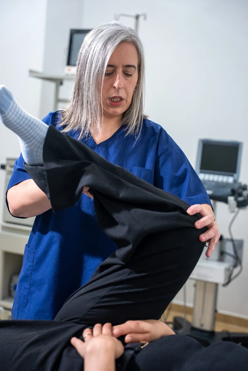
How can it present?
Groin or hip pain
Discomfort may appear in the groin, on the outside of the hip or in the buttock, especially when walking or after exertion.
Stiffness or loss of mobility
Bending, rotating or moving the leg out to the side may become difficult or painful.
Clicking or locking
Labral tears can cause clicking, catching or locking during certain movements.
Changes in gait
Pain or loss of strength can change the way you walk and how loads are distributed across the joint.
Limits on sports or daily life
Climbing stairs, getting up from a chair, putting on shoes or exercising may start to become difficult.
Your own cells, directed toward the tissue that needs treatment.
The body has natural self-repair mechanisms. However, in the face of advanced structural injuries, complex tears of the labrum or tendons, or joint impingement that causes continuous friction and microinjuries, that capacity may be insufficient.
In these cases, the contribution of cells aims to support biological processes related to tissue repair and remodeling, modulate inflammation and support the recovery of hip function.
The personalized treatment combines two key components: stem cells, part of a preparation intended to support biological processes related to tissue repair and remodeling, and immune system cells, which help direct the stem cells toward the tissue that needs treatment —cartilage, labrum, tendon or bone—.
Under the protocols of the Moviglia Method, the LYRACELL team obtains stem cells from the patient’s own fat. These are processed and expanded in the laboratory and then combined with tissue-specific immune system cells. The preparation includes pre-differentiated stem cells and immune system cells directed toward the structures identified in the diagnosis.
The combination of both cell types aims to support the body’s natural repair process, through a personalized approach and under strict safety protocols.
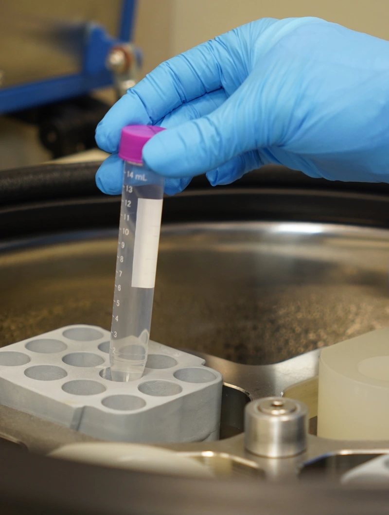
From the laboratory to your hip
The cells are processed in the laboratory and prepared according to the tissue identified in the evaluation before the targeted application.


Four stages, a process defined for each case.
From the initial evaluation to following up on your progress, each stage of treatment is defined by the medical team according to your case.
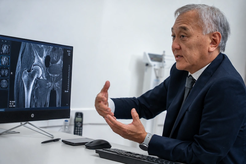
1
Evaluation
The medical history, any history of trauma or sports injuries and imaging studies —X-rays, ultrasounds, CT scans or MRIs— are analyzed to identify the affected structures and determine the degree of injury.
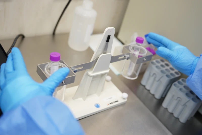
2
Treatment proposal
If the patient is eligible, a plan is designed according to the extent of the injury and the tissues involved —cartilage, labrum, bone or tendons—. Adipose tissue and peripheral blood are collected, and the cell combination adapted to the affected tissue is prepared in the laboratory.
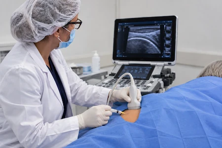
3
Application
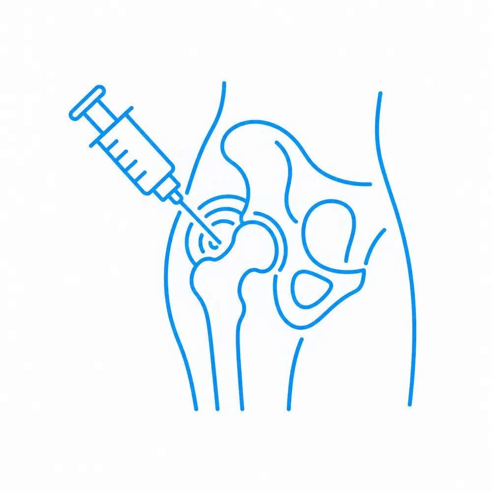
In a minimally invasive way and with ultrasound guidance, the cells are applied to the affected joint, bone or tendons. If arthroscopy is needed to smooth the surface or repair the labrum, they can be applied during or after it. The number of applications depends on the case.
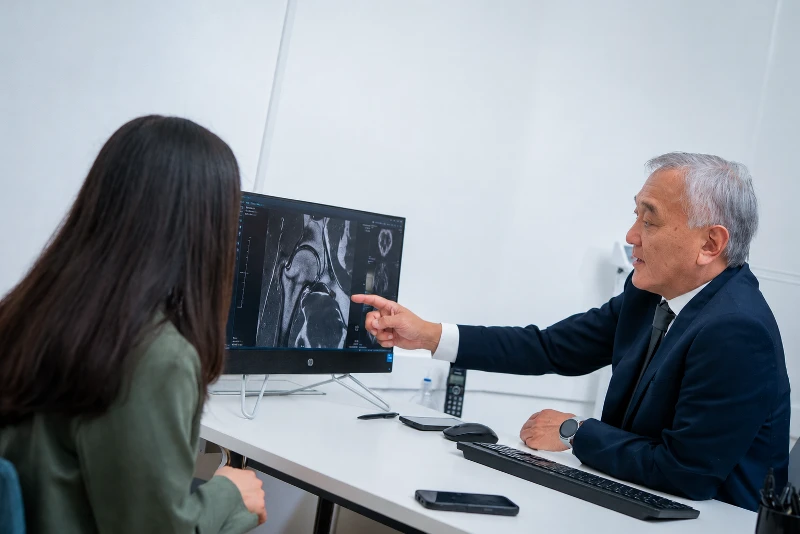
4
Follow-up
At first there may be local discomfort or inflammation. The treatment can be complemented with rehabilitation to strengthen the glutes, core and hamstrings and recover mobility. Progress is assessed at later medical check-ups.
An accurate evaluation makes it possible to treat the specific problem, not just the pain.
Could this treatment be an option for you?
The indication is defined individually based on the medical evaluation, the symptoms and imaging studies.
It may be an option if…
You have persistent pain or functional limitation in the hip.
The pain affects your walking or everyday activities.
You have lost mobility or the ability to do physical activity.
Your medical studies show a structural problem.
You want to preserve hip function and act on signs of deterioration.
Another approach may be needed if…
There is very advanced osteoarthritis, collapse of the femoral head, complete loss of joint space or continuous bone-on-bone friction. These conditions may require joint replacement.
The pain originates in the lumbar spine, such as radiculopathy or a herniated disc. In that case, the evaluation focuses on the spine.
The symptoms come from the sacroiliac joint.
The evaluation makes it possible to distinguish a structural hip problem from other causes that can produce similar symptoms.
Images that make it possible to compare progress.
Follow-up images make it possible to compare the condition of the hip before treatment and at later check-ups. They correspond to a specific case: the response, timing and degree of progress vary from patient to patient.
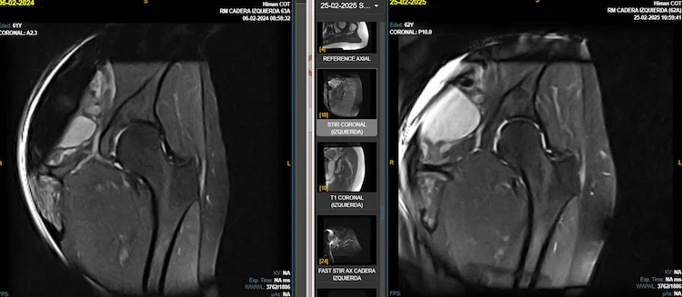
A 61-year-old patient with hip osteoarthritis. Between the two studies (February 2024 and February 2025), the patient received 3 applications of regenerative treatment in the hip. Comparing the MRIs, we found:
- More visible, continuous cartilage: the light line surrounding the head of the femur looks more defined and even in the 2025 study, indicating better coverage of the joint.
- Better joint space: the gap between the head of the femur and the pelvis looks more uniform, with better cushioning.
- Healthier-looking bone: the head of the femur looks uniform, with no signs of damage.
These results show that, one year later, this patient’s hip joint looks to be in better condition.
Results may vary from person to person. Each case is evaluated individually.
Recovering mobility also means working on function.
The recovery plan may include rehabilitation to strengthen the glutes, core and hamstrings, and gradually recover range of motion. Follow-up makes it possible to assess how hip function evolves and adapt rehabilitation to each case.
First days
Temporary local discomfort or inflammation may occur. The team indicates the necessary care and any temporary adjustments to activity and weight-bearing.
Rehabilitation and progress
Progress is assessed at later medical check-ups. Rehabilitation supports the process by strengthening the glutes, core and hamstrings, and gradually recovering joint range of motion.
Measurable results
Clinical and functional check-ups make it possible to assess pain, mobility, strength and function. When indicated, new imaging studies are added to compare progress with the starting point.
Frequently asked questions
It depends on the diagnosis. Hip osteoarthritis, femoroacetabular impingement, labral tears and tendon injuries are evaluated to determine whether cell therapy can be part of the approach.
It is one of the conditions most frequently evaluated. The indication depends on the grade of the tear, joint stability and its impact on function, and is defined after a medical evaluation.
The base treatment is autologous: the stem cells are obtained from the patient’s own adipose tissue and the immune system cells from their blood. In certain cases, depending on the medical indication, immune system cells from a selected donor may be used.
The medical team reviews your medical history, any history of trauma or sports injuries and imaging studies, such as ultrasound, X-ray or MRI.
Yes. The cells are applied with ultrasound guidance into the joint space, bone or affected tendon tissue.
In some cases it can be an alternative to surgery. If the injury requires arthroscopy to smooth the joint surface or repair the labrum, the cells can be applied during or after the procedure as a complement, not a replacement.
It depends on the type of injury and each patient’s progress. The team provides care instructions for the first days and then defines a gradual recovery plan.
The return is gradual. It depends on clinical progress, the strengthening of the glutes, core and hamstrings, and the medical team’s instructions. There is no single timeframe.
Through clinical check-ups and a functional assessment of pain, mobility and strength. When appropriate, imaging studies are also used. The results are compared with the starting point.
If the diagnosis points to another approach, the team will let you know and guide you on the next steps. For example, very advanced osteoarthritis or collapse of the femoral head may require a joint replacement. If the pain comes from the lumbar spine or the sacroiliac joint, the evaluation focuses on that area.
It is a developing field of research. Results vary depending on the type of injury and the protocol used. That is why each indication starts from an individual diagnosis and is accompanied by follow-up with measurable parameters.
The first step is understanding your case.
Every treatment begins with a medical evaluation to understand your diagnosis, identify the affected structures and determine which approach may be right for you.